Knee Muscle Anatomy Mri - Imaging Of Tumors And Tumor Like Lesions Of The Knee Sciencedirect - Use the mouse scroll wheel to move the images up and down alternatively use the tiny arrows (>>) on both side of the image to move the images.
Knee Muscle Anatomy Mri - Imaging Of Tumors And Tumor Like Lesions Of The Knee Sciencedirect - Use the mouse scroll wheel to move the images up and down alternatively use the tiny arrows (>>) on both side of the image to move the images.. Louis, usa and the rijnland hospital in leiderdorp, the netherlands. This article is based on a presentation given by david rubin and adapted for the radiology assistant by robin smithuis. The knee joint is a complex structure that involves bones, tendons, ligaments, muscles, and other structures for normal function. There is a flat area of tendon originating from the knee. The images may also help physicians to distinguish normal, healthy tissues from dead tissues(2).
These motions of the knee allow the body to perform such important movements as walking, running, kicking, and jumping. Shop your anatomē everyday essentials or add a new product to your collection. These muscles work in groups to flex extend and stabilize the knee joint. The knee joint is a complex structure that involves bones, tendons, ligaments, muscles, and other structures for normal function. T2w axial fat sat 1.
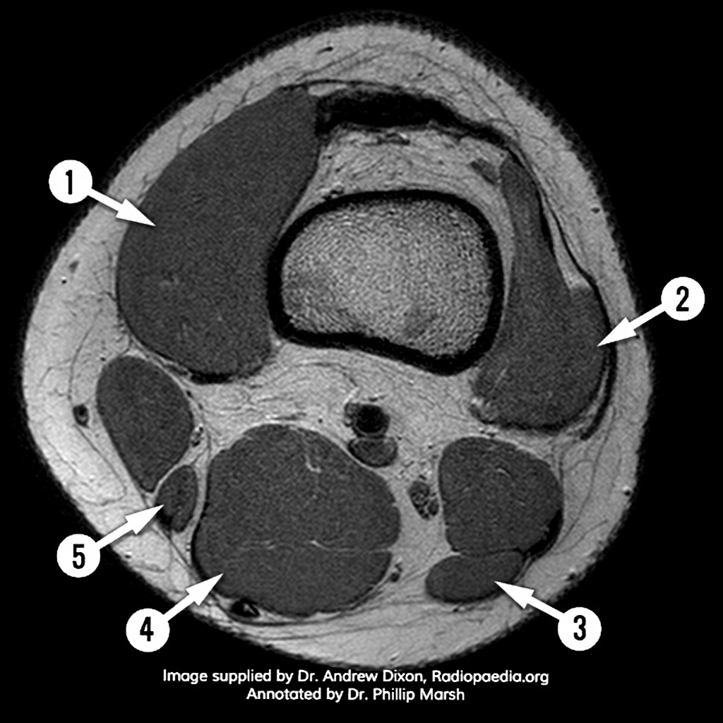
Mri Knee Axial Anatomy Quiz Radiology Case Radiopaedia Org from prod-images-static.radiopaedia.org Injuries such as anterior cruciate ligament, meniscus and rotator cuff tears are all easily diagnosed when there is a firm understanding and knowledge of human anatomy. Atlas of knee mri anatomy. Knee mri, popliteal vessels, vascular. This mri knee sagittal cross sectional anatomy tool is absolutely free to use. Medical images from an mri allow medical professionals to distinguish body tissues, including the meniscus (shock absorbers in the knee), cartilage, tendons, and ligaments. Assoc prof craig hacking and dr shu su et al. The femur, tibia and patella.the arrangement of the bones in the knee joint, along with its many ligaments, provide it with the arthrokinematics that allows for great stability, combined with great mobility.being arguably the most stressed and exposed joint of the body, the knee joint is predisposed to various. Mri knee anatomy scroll using the mouse wheel or the arrows.
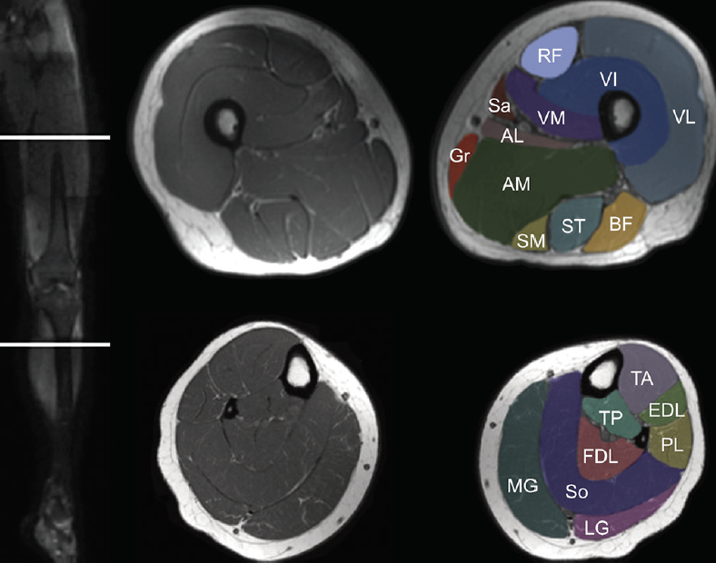
Normal mr imaging anatomy of the knee.
Coronal images are perpendicular to the long axis of the metatarsals. Louis, usa and the rijnland hospital in leiderdorp, the netherlands. Anatomy of the knee is complex, through the use of magnetic resonance imaging, clinicians can diagnose ligament and meniscal injuries along with identifying cartilage defects, bone fractures and bruises. Use the mouse scroll wheel to move the images up and down alternatively use the tiny arrows (>>) on both side of the image to move the images. In conclusion, we describe the normal mri anatomy of the distal biceps femoris and the relationship of this muscle with the common peroneal nerve. Anatomy arthrogram anatomy basic shoulder mri. Anatomy basic knee mri checklist. Shop your anatomē everyday essentials or add a new product to your collection. The knee joint is a complex structure that involves bones, tendons, ligaments, muscles, and other structures for normal function. The images may also help physicians to distinguish normal, healthy tissues from dead tissues(2). When there is damage to one of the structures that surround the knee joint, this can lead to discomfort and disability. The knee joint is a complex joint that connects three bones; Knee mri, popliteal vessels, vascular.
The images may also help physicians to distinguish normal, healthy tissues from dead tissues(2). Atlas of knee mri anatomy. Prescribe sagittal plane off axial images with line parallel to bony glenoid. Understanding the normal function of the knee joint can help you address some of these common. When there is damage to one of the structures that surround the knee joint, this can lead to discomfort and disability.
Http Www Smartview Co Wp Content Uploads 2014 02 Imagen Mr Normal Anatomia Rodilla Pdf from Mri wrist anatomy scroll using the mouse wheel or the arrows. These muscles work in groups to flex extend and stabilize the knee joint. Normal mr imaging anatomy of the knee. Medical images from an mri allow medical professionals to distinguish body tissues, including the meniscus (shock absorbers in the knee), cartilage, tendons, and ligaments. Injuries such as anterior cruciate ligament, meniscus and rotator cuff tears are all easily diagnosed when there is a firm understanding and knowledge of human anatomy. The images may also help physicians to distinguish normal, healthy tissues from dead tissues(2). Related posts of muscle anatomy knee mri muscle anatomy get body smart. Anatomy arthrogram anatomy basic shoulder mri.
In approximately 2% of the population, the anterior tibial artery branches along the keywords:
Normal mr imaging anatomy of the knee. Free access interactive and dynamic anatomical atlas saved by radiologist.ayman almatboly The femur, tibia and patella.the arrangement of the bones in the knee joint, along with its many ligaments, provide it with the arthrokinematics that allows for great stability, combined with great mobility.being arguably the most stressed and exposed joint of the body, the knee joint is predisposed to various. These muscles work in groups to flex extend and stabilize the knee joint. Magnetic resonance imaging is particularly well suited for the medical evaluation of the musculoskeletal (msk) system including the knee, shoulder, ankle, wrist and elbow. Über 7 millionen englischsprachige bücher. Injuries such as anterior cruciate ligament, meniscus and rotator cuff tears are all easily diagnosed when there is a firm understanding and knowledge of human anatomy. These muscles work in groups to flex, extend and stabilize the knee joint. In this presentation mri anatomy biceps femoris muscle. Atlas of knee mri anatomy. Thigh muscles also protect neurovascular structures as they go through the proximal hip joint to the knee and lower leg (3). The knee joint is a complex structure that involves bones, tendons, ligaments, muscles, and other structures for normal function. Medical images from an mri allow medical professionals to distinguish body tissues, including the meniscus (shock absorbers in the knee), cartilage, tendons, and ligaments.
When a muscle has different orientations of the tendons it means that there are different patterns of edema possible depending on the tendon injured. Injuries such as anterior cruciate ligament, meniscus and rotator cuff tears are all easily diagnosed when there is a firm understanding and knowledge of human anatomy. Magnetic resonance imaging (mri scan): These motions of the knee allow the body to perform such important movements as walking, running, kicking, and jumping. Magnetic resonance imaging (mri) utilizes magnet and radio waves to produce diagnostic images that allow a doctor to visualize the hips.
Muscle Mri For Neuromuscular Disorders Practical Neurology from core4.bmctoday.net Related posts of muscle anatomy knee mri muscle anatomy get body smart. Anatomy basic knee mri checklist. These muscles work in groups to flex extend and stabilize the knee joint. Anatomy muscle system 12 photos of the anatomy muscle system anatomy and physiology muscular system exam, anatomy and physiology muscular system labeling quiz, anatomy and physiology muscular system pdf, anatomy and physiology muscular system review, human anatomy muscular system quizzes, human muscles, anatomy and physiology. This article is based on a presentation given by david rubin and adapted for the radiology assistant by robin smithuis. The images may also help physicians to distinguish normal, healthy tissues from dead tissues(2). Injuries such as anterior cruciate ligament, meniscus and rotator cuff tears are all easily diagnosed when there is a firm understanding and knowledge of human anatomy. Thigh muscles also protect neurovascular structures as they go through the proximal hip joint to the knee and lower leg (3).
It is the largest synovial joint in the body and allows flexion and extension of the leg as well as some rotation in the flexed position.
Routine ankle magnetic resonance imaging (mri) tests involve taking images of the foot and ankle in the axial, coronal, and sagittal planes parallel to the tabletop(2). Normal mr imaging anatomy of the knee. Related posts of muscle anatomy knee mri muscle anatomy get body smart. Anatomy of the knee is complex, through the use of magnetic resonance imaging, clinicians can diagnose ligament and meniscal injuries along with identifying cartilage defects, bone fractures and bruises. Magnetic resonance imaging (mri) tests involve large machines that use radio wave energy pulses and a magnetic field to produce images of the shoulder (2). Three conventional mri planes that are utilized to evaluate the knee include sagittal (oblique), coronal, and transaxial planes. In approximately 2% of the population, the anterior tibial artery branches along the keywords: Doctors may recommend a knee mri if a patient experiences the following(3): Anatomy arthrogram anatomy basic shoulder mri. Coronal images are perpendicular to the long axis of the metatarsals. Related posts of knee muscle anatomy mri anatomy muscle system. The knee joint is a complex structure that involves bones, tendons, ligaments, muscles, and other structures for normal function. Anatomy arthrogram anatomy basic shoulder mri.


0 Komentar